The Exciting World of Macrocycle Structures
In this post, we discuss the unique structure of macrocyclic peptide drugs (often 4 to 24 amino acids), which falls between small-molecule drugs and biologics (such as monoclonal antibodies).
Macrocyclic peptide drug unique structure (often 4 to 24 amino acids) falls between small-molecule drugs and biologics (such as monoclonal antibodies). While linear unconstrained oligopeptides are often disordered in solution and exist in high-entropy states, cyclization reduces the available degrees of conformational freedom, facilitating binding by minimizing entropic loss. Other advantages of cyclization include higher stability against enzymatic degradation and improved affinity and specificity compared to linear peptides and small-molecule ligands. In addition, unlike biologics, macrocyclic peptides can be designed to penetrate cells and can be administered orally.
As usual, structure, of course, is essential for understanding function and the mode of action. Numerous crystal structures of complexes of macrocyclic peptide drugs with targets have been published. Malde et al. (Chem. Rev. 2019) analyzed 211 three-dimensional crystal structures of macrocyclic peptides bound to 65 different proteins. Among these, 30 were enzymes, which included 13 proteases, four kinases, and 13 other enzymes, such as hydrolases, polymerases, demethylases, ATPases, and phosphatases. The other proteins included GPCRs, glycoproteins, cell receptors, and membrane channels, among others. It should be noted that many of the analyzed proteins are available in our library of off-the-shelf structures, FastLane™ Premium and FastLane™ Standard, and can be co-crystallized with a ligand, allowing their X-ray structure to be determined within a few weeks.
The detailed analysis of macrocyclic peptide drug residues involved in stabilizing binding to proteins revealed that arginine, aspartate, glutamate, and the aromatic residues tyrosine and tryptophan are key contributors to the interactions. Polar residues such as serine, threonine, asparagine, and glutamine are also employed extensively. The aromatic residues were found to impart an amphipathic character to the macrocycle, with the side chains oriented towards the hydrophobic protein surface. Amino acids like proline and glycine help shape the macrocycle’s scaffold for optimal binding, while cysteine is frequently utilized to induce macrocyclization. The authors of the review conclude that macrocyclic peptide drugs, compared to small-molecule drugs, bind to larger and more polar solvent-exposed surfaces. The larger number of contacts can enhance the affinity and specificity of the drug for the target protein, making them more suitable for binding to the shallower, more flexible, and more polar protein-protein interfaces.
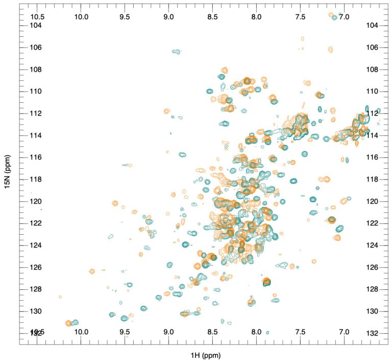
An interesting question that may arise is the relationship between the conformation of the macrocyclic peptide drug in solution and its bound state. Of the 65 complex structures analyzed by Malde et al., 15 had bound macrocycles for which an X-ray or NMR solution structure of the unbound form was reported. Among these, 10 exhibited the same or slightly altered macrocyclic peptide structure, while five displayed significant differences, suggesting protein-induced alterations in the polypeptide backbone. Since conformational restrictions reduce the energy needed for structural rearrangements into the binding state, they effectively lower the entropy penalty for binding more rigid macrocycles. This suggests that the solution state of the macrocycle could be one of the parameters to optimize during the development of the drug and needs to be verified. At SARomics Biostructures, we regularly solve X-ray crystal structures of macrocyclic peptide drug complexes for our clients. To study the conformation of peptides containing up to 30-40 amino acids in solution, NMR spectroscopy is a fast and straightforward option on modern high-field spectrometers. Our internal structure determination pipeline can also accommodate non-natural amino acids in macrocyclic peptides. Please view the details of our X-ray crystallography and NMR spectroscopy services.
Case study: The Structure of the Complexes of BamA with Inhibitors
As an illustration, we examine a recent joint publication by two teams from Genentech and PeptiDream (Sun et al., 2024). PeptiDream was founded in 2006 in Tokyo, Japan. The company focuses on discovering and developing macrocyclic peptides as next-generation peptide therapeutics and has established a proprietary drug discovery platform, PDPS (Peptide Discovery Platform System). PDPS generates diverse libraries that may contain trillions of macrocyclic peptides, which can be screened against targets of interest. The publication investigated in vitro selection methods to identify macrocyclic peptide drugs against the BamA/BAM system in Escherichia coli. BamA is the central component of the β-barrel assembly machinery (BAM), a conserved multi-subunit complex that inserts and folds the outer membrane β-barrel proteins (OMP) of Gram-negative bacteria. The BAM complex has been proposed as an antibacterial target. The C-terminal domain of BamA forms a 16-stranded β-barrel, the first and last strands of which form the so-called “lateral gate”. Earlier structural studies suggested that the observed large-scale conformational changes of the complex between open and closed states were essential for BamA function. However, which of the states is suitable for inhibitor design was unknown. Compound screening targeted various conformational states of BamA and identified two compounds (PTB1 and PTB2) that inhibited wild-type E. coli while targeting two previously unknown binding sites. To determine the molecular details of the interactions between the compounds and BamA, the structures of the PTB1-BAM and PTB2-BAM complexes were determined by cryo-EM.

The structures revealed that the PTB1 macrocycle targets a site above the lateral gate, locking BamA in a closed conformation. In contrast, the PTB2 macrocycle binds deep within the central lumen of the BamA β-barrel, indicating an open state of the lateral gate. Both compounds exhibit amphipathic characteristics, with polar/charged side chains on one side and hydrophobic/aromatic side chains on the other. PTB1 interacts with multiple extracellular loops, forming an acidic region above the closed lateral gate (see left figure below). Notably, a Zn2+ ion was coordinated by three histidine residues from PTB1 and an aspartate from the protein. The hydrophobic part of PTB1 with the aromatic side chains is inserted into the lipid environment. PTB2 also uses its amphipathic character in binding – the basic side chains engage in multiple contacts with the acidic outer opening of BamA, while the aromatic side chains are directed toward the center of the β-barrel (see figure on the right). The binding modes of PTB1 and PTB2 nicely illustrate the general observations regarding macrocycle binding made by Malde et al. mentioned above.
Follow us on LinkedIn to stay updated!


