
Protein Crystallization For X-ray Crystallography: Process & Techniques
This guide provides an overview of our protein crystallization services—from protein sample requirements to protein characterization and crystallization screening.
SARomics Biostructures combines a FastLane™ catalog of ready-to-crystallize proteins with gene-to-structure services and proximity to the fourth-generation synchrotron MAX IV, reflecting what an integrated protein crystallization service looks like at a high-level technology platform.

Overview of The General Process Of Protein Crystallization
To streamline and maximize the speed and efficiency of our services, our staff follows specific protocols throughout all crystallization and protein X-ray structure determination experiments. To ensure the quality of client samples, we have also developed general guidelines that you can download for your convenience. A product sheet for one of our crystallization-grade Protein Shop proteins shows the required quality for crystallization-grade proteins.
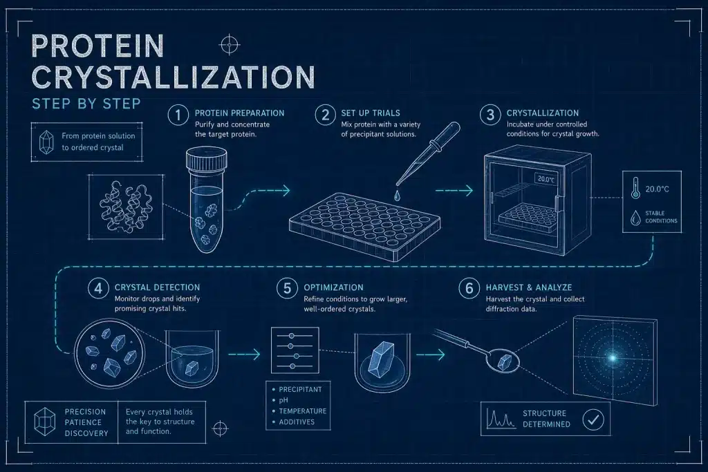
Assuming the protein has been cloned, expressed, and purified, we can begin planning the crystallization experiments. The following steps are involved in the process:
- Protein sample characterization is required to ensure the protein sample’s purity, stability, and monodispersity before setting up crystallization experiments.
- Initial screening for identification of crystallization conditions. This usually requires selecting commercial screens and assessing the protein concentration to use during screening. Commercially available plate hotels are usually used to store the crystallization plates.
- Specialized software monitors crystallization drops. We choose the most promising conditions for further optimization.
Optimization of positive hits. We usually use in-house-designed screens to further optimize crystallization conditions. - The final step involves crystal preparation, crystal testing, and X-ray data collection.
The image gives a schematic presentation of the protein crystallization process. Below, we discuss some details of the different stages. On our educational site, you may find more information on the basic principles of protein crystallization.
1. Protein Sample Characterization
The analysis of a protein-ligand complex structure and the interpretation of the results depend heavily on the quality and resolution of the X-ray data, which in turn depend on the quality of the crystals used for X-ray data collection. As a rule of thumb, a resolution of 2.4 Å or better is considered best for ligand-complex characterization. It should also be noted that a substantial fraction of crystallization failures stems from poor sample quality before a crystallization drop is set. For this reason, to assess protein sample quality before running crystallization screens, we always begin with quality control (QC). The controls aim to remove factors that can prevent protein crystallization or result in poor-quality crystals and, subsequently, poor diffraction quality. Such factors include high flexibility in certain regions of the protein (typically surface loop regions), instability (and partial degradation) in the crystallization buffer at a given temperature, polydispersity (the presence of aggregated material or different oligomeric forms of the protein), and time-dependent denaturation. The importance of QC cannot be overstated and is essential to accelerate the project and prevent the loss of precious material and time in crystallization trials that yield no crystals.
As a crystallization services provider, we strictly follow predefined protocols that standardize settings and procedures to ensure consistency, reproducibility, and reliability.
After expressing and purifying the protein, or upon receiving it from a client, we use various biophysical techniques to assess sample quality.
- We evaluate the protein’s state in solution, including aggregates, denatured material, polydispersity, and impurities.
- We assess stability across various buffers, pH values, and salt concentrations commonly used in crystallization screening.
- We verify the protein’s stability at room temperature and in cold storage, since crystallization trials are typically conducted at two temperatures.
We use the following biophysical techniques for sample characterization:
- Differential scanning fluorimetry (DSF) assesses protein stability across different buffers and temperatures.
- Size-exclusion chromatography (SEC) is often required to remove potential aggregates.
- Dynamic light scattering (DLS) and mass spectrometry (MS) assess sample monodispersity.
- For additional quality control and to detect partially unfolded regions or regions of high flexibility, sometimes NMR spectroscopy is used via HSQC fingerprint analysis.
2. Crystallization Screens, Initial Hit Identification
Screening and optimizing crystallization conditions for a new protein can take as little as 3-6 weeks, though some samples may take longer. Please note that the process is substantially accelerated if the protein is found in our FastLane™ libraries. The libraries contain 600+ proteins ready for crystallization (Premium list) or for expression, purification, and crystallization according to verified protocols (Standard list). However, even when the protein’s structure is known from previous studies, it is often necessary to perform new screening experiments to obtain high-quality crystals. Slight changes in expression, purification, or characterization protocols, or the presence of new additives in the buffer, such as ligands for co-crystallization to obtain a protein-ligand complex, may also require modifications to the crystallization conditions. In addition, some experiments, such as crystallographic fragment screening, require hundreds of stable crystals with well-reproducible crystallization conditions.
We initially run hundreds of screens using commercial sparse-matrix screens such as those available at Hampton Research. We routinely use high-throughput, high-precision liquid-handling robotics for the initial screens and during subsequent optimization of newly identified crystallization conditions. Typically, approximately 5 mg of protein at a concentration of around 10 mg/ml is required at the initial stage. Lower protein concentrations can be used if solubility is an issue. Posible temperature effects are tested using temperature screening that typically starts at 4°C and 20°C as initial reference points.
3. Optimization Of Initial Hits, Ligand Complex Crystallization
The initial crystallization conditions identified during screening almost always require further optimization. Well-formed crystals typically range from 0.5 to 2 mm in size, depending on the protein and crystallization conditions. During optimization, parameters such as buffer pH, salt concentration, additive concentration, and even the precipitant type may need adjustment to produce crystals of the required size. We may also need to adjust drop volume to optimize crystal size and explore alternative temperatures during this stage. Volume and temperature variations may potentially affect crystal nucleation and growth rates, improving crystal quality and yield (e.g., for crystallographic fragment screening). We use automated, temperature-controlled plate hotels designed for high-throughput protein crystallization and monitoring. We monitor crystallization plates using an imaging system, such as the Formulatrix Rock Imager.
Crystallization of protein-ligand complexes is extensively used in structure-based drug design (SBDD). Co-crystallization with a ligand of interest often begins after establishing crystallization conditions for the apo-protein. There are two primary methods for obtaining a ligand complex: (1) adding a ligand solution to the crystallization drop containing crystals and soaking the crystals with the ligand, and (2) co-crystallization with the ligand. Soaking may work because protein crystals contain a relatively high percentage of solvent (crystallization buffer), allowing the ligand to diffuse into the crystal. If the ligand-binding site within the crystal is exposed and accessible to solvent, the ligand will bind. Conversely, if the site is deeply buried within the protein or obscured by crystal contacts, it becomes inaccessible to ligands.
In many cases, ligand soaking cracks the crystal, resulting in poor X-ray diffraction. In such cases, we use co-crystallization with a ligand, which can be applied in different ways. The simplest approach is to mix the ligand solution with the protein before preparing crystallization drops. However, ligand binding may trigger a conformational transition in the protein, potentially requiring reoptimization of crystallization conditions. In some cases, co-crystallization may be combined with seeding, which may help in obtaining crystals of the complex. We also need to note that, in all these experiments, ligand affinity is an important factor to consider.
More information, with detailed discussion and guidelines for the successful generation of protein–ligand complex crystals, can be found in this excellent publication. You can also find some practical details on the Hampton Research website.
4- Cryoprotection and Synchrotron Data Collection
Before a crystal can be flash-cooled in liquid nitrogen for transport and data collection, it must be briefly transferred to a cryoprotectant solution containing glycerol, ethylene glycol, or a high-concentration PEG. The goal is vitrification: replacing the solvent in the crystal with a glass-like solid that avoids forming crystalline ice during cooling. Crystalline ice results in poor diffraction quality. The challenge is that the cryoprotectant must not disrupt protein packing, and both concentration and soaking time must be tested empirically for each crystal system. For fragile crystals, stepwise transfer into increasing cryoprotectant concentrations is safer than a direct jump to full concentration. The crystal is inspected after each transfer step; cracking, opacity, or dissolution indicates an incompatible cryoprotectant or an excessive concentration. Once a cryosolution tolerated by the crystal is found, excess liquid is removed from around the mounted loop, and the crystal is immediately plunged into liquid nitrogen.
At the synchrotron, data collection is relatively fast, often minutes per crystal. As mentioned above, dataset quality largely depends on the earlier stages: the crystal’s quality (internal order) and the quality of the cryoprotection. Direct access to a high-brilliance synchrotron source, like MAX IV Laboratory in Lund, is a clear advantage because it removes the need to send crystals to remote synchrotrons. This is also essential for crystallographic fragment screening, as in this case a large number of crystals are grown directly at the beamline.